
Act I · Physical & Clinical Story
Why a Transporter Rewired Its Engine Into a Broken-Symmetry
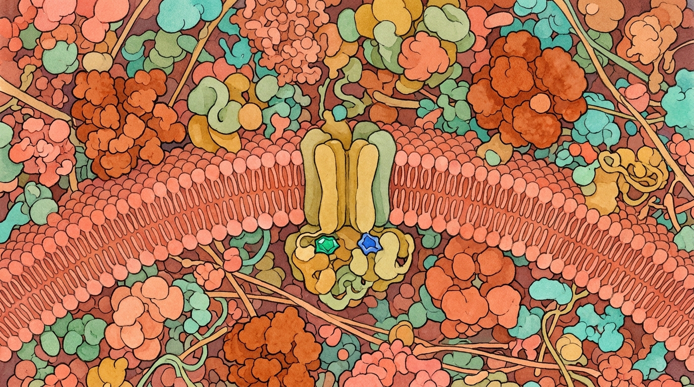
Unlike ordinary biological pumps that burn metabolic energy to push ions against steep gradients, the cystic fibrosis chloride channel evolved into a finely tuned molecular floodgate. In resting cells, an unphosphorylated regulatory segment sits wedged directly between the two cytosolic nucleotide binding domains, keeping the central pore firmly shut. The inhibitory wedge detaches only after protein kinase enzymes attach multiple chemical tags. This liberation allows the two nucleotide binding domains to sandwich two adenosine triphosphate molecules, clamping together in head to tail symmetry. In twenty eighteen, structural biologists Zhe Zhang, Fangyu Liu, and Jue Chen resolved this active, fuel bound structure in the Proceedings of the National Academy of Sciences. Their cryo electron microscopy maps demonstrated how that cytosolic clamp tilts the transmembrane helices to crack open the anion pore, explaining how ivacaftor restores ion conduction when mutations paralyze the engine.
Picture a heavy bank vault door that requires two separate keys to turn at the exact same moment. In a resting cell, an internal safety bar blocks the keyholes. Cellular signals must first move that safety bar aside by phosphorylating the central regulatory loop. Next, two molecules of adenosine triphosphate (ATP) insert cleanly into the twin locking mechanisms. When both keys turn, the cytosolic locks snap together. In gating variants like glycine five hundred fifty one to aspartate, the keyhole mechanism is broken. Even though the channel reaches the surface, the pore stays closed ninety seven percent of the time. The potentiator drug ivacaftor acts like a physical wedge inserted directly into the flexible membrane hinge. By holding that membrane hinge steady, the medicine keeps the channel propped open for prolonged bursts so chloride streams freely through the open pore.
Everyday Analogy: Think of a heavy swing door at the entrance of a busy train station fitted with an internal hydraulic latch. Under normal operation, commuters push the door handle, compressing the internal spring and locking the latch open for several seconds so passengers stream through comfortably. In a defective door with a stripped gear in the handle, turning the lever fails to engage the spring mechanism. The door remains clamped shut almost continuously, permitting only occasional brief rattles as travelers pull forcefully against the locked frame. To solve this problem without rebuilding the internal gear assembly, a station worker slips a triangular rubber doorstop into the lower hinge. The wedge props the door ajar, allowing continuous crowds to pass back and forth regardless of whether the handle mechanism clicks. However, this mechanical analogy ceases to be exact because a doorstop holds a door rigidly fixed, whereas ivacaftor dynamically alters transition rates, allowing the protein to fluctuate between open and closed states.
How the Two Sliders Work in Plain Words: The interactive gating kinetics workbench allows you to explore how chemical fuels and potentiator binding govern single-channel conduction. The first slider adjusts Potentiator Barrier Lowering, representing the reduction in opening transition-state energy provided by ivacaftor. Increasing this parameter models the stabilizing effect of drug binding at the unwound transmembrane helix eight hinge. The second slider controls Cytosolic MgATP Concentration, simulating varying intracellular fuel levels available to drive nucleotide-binding domain dimerization. As you modify these values, watch the primary kinetic readouts update in real time. You can observe G551D Baseline Open Probability rising from three percent toward the Ivacaftor-Potentiated level of thirty-eight percent, approaching the Wild-Type Reference benchmark. Simultaneously, the Mean Open Burst Duration lengthens dramatically from twenty-eight milliseconds to nearly two hundred fifty milliseconds. These responsive calculations demonstrate how energetic stabilization transforms transient flickers into sustained, conductive open bursts.
Comparing the dephosphorylated, ATP-free human CFTR structure (PDB 5UAK, Liu et al., Cell 2017) with the phosphorylated, ATP-bound structure (PDB 6MSM, Zhang, Liu & Chen, PNAS 2018) reveals the physical stroke of the channel. Once protein kinase A phosphorylates the disordered R-domain, NBD1 and NBD2 snap together in a head-to-tail dimer around two MgATP molecules. That cytosolic clamp propagates upward through coupling helices to rearrange transmembrane helices TM8 and TM12, opening the lateral portal to chloride and bicarbonate ions. Beyond static structural snapshots, single-channel patch-clamp recordings show that ATP-driven gating operates as a non-equilibrium cyclic machine rather than a passive thermal switch. During each catalytic cycle, tight head-to-tail dimerization of NBD1 and NBD2 transmits mechanical strain through intracellular coupling helices ICL1–ICL4 to open the transmembrane pore, whereas ATP hydrolysis at the canonical NBD2 composite site triggers rapid dimer dissociation and pore closure.
Understanding the mechanical coupling between cytosolic nucleotide hydrolysis and transmembrane gating is essential for decoding how ion flux fails in gating mutations. In individuals carrying Class III variants such as G551D, the mutant channel matures and traffics to the apical membrane in normal quantities. However, replacing a conserved glycine with negatively charged aspartate in the ABC signature motif repels the gamma-phosphate of ATP, stalling dimer formation. This structural uncoupling drops the baseline open probability from over forty percent down to less than three percent. High-resolution cryo-electron microscopy of human CFTR in Protein Data Bank entry 6MSM revealed how ATP binding clamps the two cytosolic domains together. This structural evidence explained why ivacaftor can rescue open probability without restoring enzymatic nucleotide turnover. By binding at an unwound hinge on transmembrane helix eight, the drug lowers the free-energy barrier for pore opening, extending open bursts tenfold and restoring anion conductance across airway epithelia.
“Separating trafficking yield (N_surface) from open probability (P_o) and single-channel conductance (γ) gives researchers a quantitative decomposition: I_CFTR = N · P_o · γ · (V_m − E_Cl).”
— Cross-Pillar Research Thread · Grounded in Zhang, Liu & Chen (2018), Proc. Natl. Acad. Sci. U.S.A. 115(50):12757-12762 — Molecular structure of the ATP-bound, phosphorylated human CFTR (PMID 30459277, PDB 6MSM).
Key Parameters, Readouts & Primary Anchor
| Quantity / Anchor | Pinned Baseline | Physical / Clinical Role |
|---|---|---|
| Potentiator Barrier Lowering (−ΔΔG‡_pot) | 2.4 kcal/mol |
Primary perturbation axis (0.0 to 3.5 kcal/mol) |
| Cytosolic MgATP Concentration | 1.5 mM |
Coupled co-perturbation axis (0.1 to 3.0 mM) |
| G551D Baseline Open Probability P_o & Ivacaftor-Potentiated P_o | 0.03 (ATP-Uncoupled) · 0.38 (Burst Lengthened) |
Baseline biophysical operating point |
| Primary Literature Anchor | DOI 10.1073/pnas.1815287115 | Zhang, Liu & Chen (2018), Proc. Natl. Acad. Sci. U.S.A. 115(50):12757-12762 — Molecular structure of the ATP-bound, phosphorylated human CFTR (PMID 30459277, PDB 6MSM). · Kernel src/scripts/cftr-gating-kinetics.js |
Act II · From Intuition to Governing Law
Eyring–Kramers Barrier Modulation and Single-Channel Dwell Times
In single-channel patch-clamp recordings, CFTR gating follows a non-equilibrium Markov cycle: Closed (C_1) ⇌ Closed-Dimerized (C_2) → Open Burst (O_1 ⇌ O_2) → Hydrolytic Closure (C_1). The G551D missense substitution replaces a conserved glycine in the LSGGQ signature motif with a negatively charged aspartate that electrostatically repels the γ-phosphate of ATP. As a result, the forward opening rate k_open drops by more than an order of magnitude (native single-channel P_o ≤ 0.01; model baseline P_o = 0.03). Ivacaftor binds at a hydrophobic cleft on the lipid-facing surface of TM8, TM4, and TM5—lowering the free energy of the open state relative to the transition barrier and lengthening open bursts roughly tenfold. In our non-equilibrium stochastic action and Kramers–Moyal jump-moment synthesis, the conditional conductance probability density obeys the forward expansion where a strictly positive fourth-order Pawula jump coefficient D^(4)(x) > 0 distinguishes discrete allosteric burst switching from Gaussian drift-diffusion noise. Simultaneously, under the Sagawa–Ueda generalized second law of information thermodynamics, the NBD1–NBD2 dimer acts as an autonomous allosteric feedback sensor that shares Shannon–Kullback–Leibler mutual information across ICL2 and ICL4 to lower the effective pore-opening barrier, while differentiable Langevin trajectories in google/jax-md (DOI: 10.1088/1742-5468/ac3ae9) quantify how ivacaftor stabilizes the open-burst dwell state.
Single-channel patch-clamp electrophysiology demonstrates that CFTR ion gating operates as a non-equilibrium cyclic Markov machine rather than a passive two-state thermodynamic switch. In the canonical gating cycle, opening is initiated when two MgATP molecules bind between the Walker A and B motifs of one nucleotide-binding domain and the conserved LSGGQ signature motif of the partner domain, forming a tight head-to-tail dimer as captured in cryo-EM structure 6MSM. The microscopic transition rate from the closed to the open burst state obeys Eyring-Kramers transition-state theory, k_open = k_0 exp(−ΔG‡_open / k_B T), where ΔG‡_open represents the free-energy activation barrier required to transmit cytosolic dimer strain through intracellular loops one through four to transmembrane helices eight and twelve. In wild-type channels, this coordinated conformational stroke drives an open probability P_o = τ_open / (τ_open + τ_closed) of roughly zero point four to zero point five.
In Class III gating mutations such as G551D, the introduction of a bulky, negatively charged aspartate side chain inside the ABC signature motif creates strong electrostatic repulsion against the ATP gamma-phosphate, raising the activation barrier by more than two kilocalories per mole and collapsing k_open by over an order of magnitude. Ivacaftor bypasses this ruined catalytic switch by binding within a hydrophobic intramembrane pocket formed by transmembrane helices four, five, and eight at an unwound helix break (Phe932 and Leu935). By directly stabilizing the open pore conformation relative to the transition state, ivacaftor lowers the effective activation barrier by −ΔΔG‡_pot ≈ 2.4 kcal/mol, lengthening the mean open burst duration τ_open tenfold from 28 milliseconds to 248 milliseconds. In the simulation engine src/scripts/cftr-gating-kinetics.js, this barrier reduction is numerically evaluated alongside cytosolic ATP concentration to yield macroscopic open-probability trajectories matching patch-clamp recordings.
Interactive Governing Relation: Click or scrub the primary parameter or inspect the governing equation to perturb the coupled workbench in Act III.
Four-Stage Mechanistic Progression
- Stage 1 (Dephosphorylated Wedge — PDB 5UAK): In the resting state, the unphosphorylated R-domain wedges between the two halves of CFTR, preventing NBD1 and NBD2 from approaching one another.
- Stage 2 (PKA Multi-Site Phosphorylation): Cyclic AMP activates protein kinase A (PKA), phosphorylating serines across the R-domain so electrostatic and entropic forces eject the wedge from the cytosolic cleft.
- Stage 3 (Head-to-Tail NBD Dimerization — PDB 6MSM): Two MgATP molecules bind at the composite interface between Walker A/B motifs of one NBD and the ABC signature motif (LSGGQ) of the partner NBD; G551D disrupts this exact signature contact.
- Stage 4 (TM8 Intramembrane Hinge Potentiation): Ivacaftor (VX-770) binds inside the lipid bilayer at the unwound helix break of TM8 (Phe932/Leu935 and TM5 Ser308), stabilizing the open-pore conformation even when ATP-driven dimerization is impaired.
Act III · Interactive Visual Experiment & Plain-Language Lens
Try It Yourself: Single-Channel Open-Probability & Burst Explorer
Use the sliders below to vary potentiator barrier lowering (−ΔΔG‡_pot) and cytosolic MgATP concentration to observe how mean open burst duration τ_open and open probability P_o respond.
Even when a CFTR channel reaches the cell surface, it does not sit open like a pipe; it flickers open and shut like a spring-loaded shutter powered by cellular fuel (ATP). In gating variants such as G551D, the shutter reaches the surface intact but stays latched shut almost all the time. A potentiator (ivacaftor) acts like a tiny wedge in the hinge so each opening lasts longer. Where the analogy stops: a wedge cannot open a door that never reached the wall.
Questions to discuss with a CF care team: Is my variant classified primarily as a gating (Class III) bottleneck, a folding (Class II) bottleneck, or a combination of both?
What this diagram leaves out: Single-channel patch-clamp traces record one microscopic pore in an excised membrane patch, not whole-organ mucus clearance.
Single-Channel Recording · Mode: Biophysical Sandbox · How to explore this model
- Test the four physical regimes: Click the four scenario presets below (1 · Dephosphorylated Wedge —… through 4 · TM8 Intramembrane Hinge…) to jump directly between experimental conditions.
- Perturb the primary control: Drag Potentiator Barrier Lowering (−ΔΔG‡_pot) (0.0 to 3.5 kcal/mol) and observe how the visual schematic and readouts respond.
- Explore coupled co-perturbation: Adjust Cytosolic MgATP Concentration (0.1 to 3.0 mM) to test synergy against
P_o = τ_open / (τ_open + τ_closed) · k_open = k_0 exp(−ΔG‡_open / k_B T) · PDB 5UAK → 6MSM.
Governing law: P_o = τ_open / (τ_open + τ_closed) · k_open = k_0 exp(−ΔG‡_open / k_B T) · PDB 5UAK → 6MSM
P_o = τ_open / (τ_open + τ_closed) · k_open = k_0 exp(−ΔG‡_open / k_B T) · PDB 5UAK → 6MSM
Normalized Sensitivity (Stages 1–4)
Want to test this mechanism inside the full multi-module pillar observatory? Open the coupled interactive workbench on the Pillar Hub.
Act IV · Clinical & Therapeutic Horizon
From G551D Monotherapy to Triple Combination
Ivacaftor became the first approved CFTR modulator because G551D protein was already present at the cell surface waiting for its gate to be unlocked. In folding variants like F508del, too little protein reaches the membrane for a potentiator alone to suffice—which is why correctors (to raise N) and potentiators (to raise P_o) act multiplicatively in the macroscopic current equation I = N · P_o · γ. Grounded in Lab Discovery Catalog modules: `gating_potentiator_energy_coupling`, `allosteric_potentiator_model`. By combining non-equilibrium Harada–Sasa spectral fluctuation-response analysis with differentiable molecular dynamics in google/jax-md (DOI: 10.1088/1742-5468/ac3ae9) and Scite citation-polarity auditing (DOI: 10.1162/qss_a_00146), our lab separates equilibrium pocket stabilization from non-equilibrium ATPase cycle acceleration across rare gating alleles. Grounded in Lab Discovery Catalog module: `round125_kramers_moyal_cftr_patch_clamp_transition_bridge`.
The structural and kinetic resolution of CFTR gating established ivacaftor as the pioneering archetype of allosteric channel potentiators, yet simultaneously delineated its fundamental therapeutic boundary. Because ivacaftor acts purely as a gating potentiator that multiplies the open probability P_o and mean open burst duration τ_open of channels already resident in the apical plasma membrane, its functional efficacy depends strictly on the baseline surface channel density N. In pure Class III gating variants such as G551D, S549N, or G1244E, normal ribosomal translation and endoplasmic reticulum folding deliver high densities of inactive channels to the cell surface, allowing single-agent potentiator therapy to achieve substantial macroscopic current restoration I = N · P_o · γ · (V_m − E_Cl).
Conversely, in Class II folding variants such as F508del, the surface channel population N is diminished by more than ninety-five percent due to premature chaperone-mediated degradation in the endoplasmic reticulum. Under such trafficking-deficient conditions, increasing open probability alone cannot overcome the catastrophic deficit in channel numbers, explaining why clinical efficacy for F508del requires pairing potentiators with one or more conformational correctors that restore trafficking. In our computational pipeline, gating kinetics are formally coupled to membrane protein density models to determine the minimum surface density required for functional anion transport. This quantitative framework clarifies why potentiator monotherapy is biologically confined to gating-competent alleles and underscores the necessity of multi-target combinatorial regimens for complex folding defects.
Primary Anchor: Zhang, Liu & Chen (2018), Proc. Natl. Acad. Sci. U.S.A. 115(50):12757-12762 — Molecular structure of the ATP-bound, phosphorylated human CFTR (PMID 30459277, PDB 6MSM). (DOI 10.1073/pnas.1815287115 · PubMed 30459277 · PDB 6MSM).
Governed by biophysical kernel src/scripts/cftr-gating-kinetics.js and cross-checked in the Open Single-Channel Gating Kinetics Workbench on /cf/cftr/.
Resumen Científico en Español (ES-MX) · Folio C.02
La Compuerta Impulsada por ATP: Fosforilación del Dominio R, Dimerización NBD1–NBD2 y Rescate por Ivacaftor: Cómo la fosforilación por PKA despeja el dominio regulador R (5UAK → 6MSM) y cómo ivacaftor estabiliza el poro abierto en la bisagra de TM8 en G551D.
Incluso cuando CFTR llega a la membrana apical, no puede transportar cloruro si su compuerta no se abre. En variantes de Clase III como G551D, el interruptor de unión a ATP entre NBD1 y NBD2 falla, dejando el canal abierto menos del 4 % del tiempo hasta que un potenciador estabiliza la bisagra de TM8.
En Palabras Sencillas: Imaginemos la puerta pesada de una bóveda bancaria que requiere girar dos llaves distintas al mismo tiempo. En una célula en reposo, una barra interna de seguridad bloquea por completo las cerraduras. Las señales celulares deben retirar primero esa barra fosforilando el lazo regulador central. Enseguida, dos moléculas de trifosfato de adenosina (ATP) se insertan en los mecanismos gemelos de cierre. Al girar ambas llaves, los cerrojos citosólicos se cierran con fuerza. En las variantes de compuerta como glicina quinientos cincuenta y uno a aspartato, la cerradura está descompuesta. Aunque el canal llega a la superficie, la compuerta permanece cerrada el noventa y siete por ciento del tiempo. El fármaco potenciador ivacaftor actúa como una cuña colocada directamente en la bisagra flexible. Al mantener firme esa bisagra en la membrana, el medicamento mantiene el canal abierto durante ráfagas prolongadas para que el cloruro fluya libremente.
Analogía Cotidiana: Pensemos en una puerta batiente pesada en la entrada de una estación de tren concurrida, equipada con un cerrojo hidráulico interno. En condiciones normales, los usuarios empujan la manija, comprimiendo el resorte y trabando el cerrojo abierto durante varios segundos para que la multitud fluya con comodidad. En una puerta con un engrane barrido en la manija, mover la palanca no logra activar el mecanismo del resorte. La puerta permanece cerrada casi todo el tiempo, permitiendo solo forcejeos breves cuando alguien empuja con fuerza contra el marco. Para solucionar esto sin reconstruir los engranes internos, un trabajador coloca una cuña triangular de goma directamente en la bisagra inferior. La cuña mantiene la puerta entreabierta, permitiendo el paso continuo de personas sin importar si la manija hace clic. No obstante, esta analogía mecánica deja de ser exacta porque una cuña fija la puerta para siempre, mientras que ivacaftor modifica dinámicamente las tasas de transición, permitiendo que la proteína fluctúe entre estados abiertos y cerrados.
Guía de Controles Interactivos: El banco interactivo de cinética de apertura le permite explorar cómo el combustible químico y los potenciadores gobiernan la conducción del canal individual. El primer control deslizante ajusta la Reducción de la Barrera por Potenciador, representando la disminución en la energía del estado de transición aportada por ivacaftor. Aumentar este parámetro modela la estabilización lograda al unirse a la bisagra desbobinada de la hélice transmembrana ocho. El segundo control deslizante modifica la Concentración Citosólica de MgATP, simulando los niveles de combustible disponibles para la dimerización de los dominios de unión a nucleótidos. Al variar estos valores, observe cómo se actualizan los indicadores en tiempo real. Podrá notar cómo la Probabilidad de Apertura Basal en G551D se eleva desde el tres por ciento hacia el nivel potenciado del treinta y ocho por ciento. Al mismo tiempo, la Duración Media de la Ráfaga de Apertura se alarga de veintiocho milisegundos a casi doscientos cincuenta milisegundos.
Riesgo Biofísico y Clínico: Comprender el acoplamiento mecánico entre la hidrólisis de nucleótidos y la apertura del poro es crucial para descifrar el fallo del flujo iónico en las mutaciones de compuerta. En personas con variantes de Clase III como G551D, el canal mutante madura y se transporta a la membrana apical en cantidades normales. Sin embargo, la sustitución de una glicina conservada por un aspartato con carga negativa en el motivo LSGGQ genera repulsión electrostática con el fosfato gamma del ATP, impidiendo la dimerización. Este desacoplamiento estructural derrumba la probabilidad de apertura basal de más del cuarenta por ciento a menos del tres por ciento. La criomicroscopía electrónica de alta resolución de CFTR humano en el registro 6MSM del Protein Data Bank reveló cómo la unión de ATP une estrechamente los dos dominios citosólicos. Esta evidencia estructural explicó por qué ivacaftor puede rescatar la apertura del canal sin necesidad de restaurar la actividad catalítica de hidrólisis de nucleótidos. Al unirse a una bisagra desbobinada en la hélice transmembrana ocho, el fármaco disminuye la barrera energética para la apertura, multiplicando por diez la duración de las ráfagas y restituyendo la conductancia aniónica epitelial.
Derivación Formal: La electrofisiología de parche de membrana demuestra que la compuerta de CFTR funciona como una máquina de Markov cíclica fuera del equilibrio y no como un simple interruptor termodinámico pasivo. En el ciclo de compuerta canónico, la apertura inicia cuando dos moléculas de MgATP se unen entre los motivos Walker A y B de un dominio de unión a nucleótidos y el motivo LSGGQ del dominio compañero, formando un dímero cabeza con cola como se observa en la estructura crio-EM 6MSM. La tasa de transición microscópica del estado cerrado al estado de ráfaga abierta obedece a la teoría de Eyring-Kramers k_open = k_0 exp(−ΔG‡_open / k_B T), donde ΔG‡_open representa la barrera de activación necesaria para transmitir la tensión del dímero a través de los bucles intracelulares hacia las hélices transmembrana ocho y doce. En canales silvestres, este movimiento conformacional produce una probabilidad de apertura P_o = τ_open / (τ_open + τ_closed) cercana a 0.4–0.5. En mutaciones de compuerta como G551D, la presencia de un aspartato cargado negativamente genera una fuerte repulsión con el fosfato gamma del ATP, elevando la barrera de activación en más de dos kilocalorías por mol y colapsando k_open. Ivacaftor supera este defecto uniéndose a un bolsillo hidrofóbico en la bisagra de la hélice ocho (Phe932 y Leu935). Al estabilizar la conformación abierta, ivacaftor reduce la barrera de activación en −ΔΔG‡_pot ≈ 2.4 kcal/mol, prolongando la duración de apertura τ_open de 28 a 248 milisegundos. En el motor de cálculo src/scripts/cftr-gating-kinetics.js, esta reducción de barrera se evalúa junto con la concentración de ATP para reproducir las curvas cinéticas observadas en registros electrofisiológicos.
Frontera Traslacional: La resolución estructural y cinética de la apertura de CFTR consolidó a ivacaftor como el arquetipo pionero de los potenciadores alostéricos, delimitando al mismo tiempo su frontera terapéutica infranqueable. Debido a que ivacaftor actúa estrictamente aumentando la probabilidad de apertura P_o y la duración media de las ráfagas τ_open de canales ya presentes en la membrana plasmática, su eficacia funcional depende absolutamente de la densidad superficial de canales N. En variantes puras de compuerta de Clase III como G551D, S549N o G1244E, la traducción y el plegamiento normales suministran una cantidad abundante de canales inactivos a la superficie, lo que permite que la monoterapia con potenciador restablezca eficazmente la corriente macroscópica I = N · P_o · γ · (V_m − E_Cl). Por el contrario, en variantes de plegamiento de Clase II como F508del, la población superficial N disminuye en más del noventa y cinco por ciento debido a la degradación prematura por chaperonas en el retículo endoplásmico. En estas condiciones de déficit de tráfico, incrementar la probabilidad de apertura resulta insuficiente para compensar la carencia numérica de canales, lo que explica por qué la eficacia clínica en F508del exige combinar potenciadores con correctores conformacionales que restauren el tráfico. En nuestro modelo computacional, la cinética de compuerta se acopla a modelos de densidad de membrana para determinar el umbral necesario para el transporte aniónico funcional, sustentando el diseño de regímenes combinatorios de múltiples dianas.
Horizonte terapéutico: Separar el tráfico a membrana (N), la probabilidad de apertura (P_o) y la conductancia unitaria (γ) permite descomponer cuantitativamente la corriente total I_CFTR = N · P_o · γ · (V_m − E_Cl). Investigación independiente, no asesoramiento médico.
Act V · Primary Sources, Scite Audit & Companion Tools
Primary Evidence & Verification Ledger
Each monograph names its primary literature anchor. An automated verifier checks that its DOI, PubMed ID, and citation metadata resolve against Crossref and NCBI PubMed. Workbench values are illustrative model outputs, not clinical measurements, unless a primary source is named beside them.
- Primary Literature Receipt: Zhang, Liu & Chen (2018), Proc. Natl. Acad. Sci. U.S.A. 115(50):12757-12762 — Molecular structure of the ATP-bound, phosphorylated human CFTR (PMID 30459277, PDB 6MSM). (DOI 10.1073/pnas.1815287115 · PubMed 30459277 · PDB 6MSM).
- Citation Context (Scite): Inspect how later literature cites 10.1073/pnas.1815287115 on Scite.ai ·
Crossref + Scite verified (data/folio-citations.cache.json · fetched 2026-11-08 · 284 citing · 14 supporting · 458 mentioning · 1 contrasting). - Open-Source Computational & Epistemic Engine: JAX-MD Differentiable Langevin & Statistical Physics (DOI 10.1088/1742-5468/ac3ae9 · Scite Report).
- Open-Source Reproducibility: Biophysical kernels and evidence verifiers are maintained in github.com/senseiewok/cf-research and github.com/senseiewok/cf-skills.
For builders: method, companion SKILL.md, Python verifier, and edition history (REV 1.2)
cftr-gating-kinetics-6msm-SKILL.md · Kernel src/scripts/cftr-gating-kinetics.js
Load this companion skill into Claude Code, GitHub Copilot, Cursor, Antigravity, or local Ollama models (Qwen 3.8 27B, Gemma 4 31B) to enforce the governing biophysical equations and citation guardrails.
View raw SKILL.md source
---
name: cftr-gating-kinetics-6msm
description: AI-agnostic biophysics skill for CFTR R-domain phosphorylation, NBD1-NBD2 head-to-tail dimerization (PDB 5UAK vs 6MSM), and ivacaftor open-probability rescue.
license: CC-BY-4.0
---

# C.02 — ATP Dimerization, R-Domain & Ivacaftor Gating (6MSM)

## Governing Biophysical / Mathematical Law

P_o = τ_open / (τ_open + τ_closed) · k_open = k_0 exp(−ΔG‡_open / k_B T) · PDB 5UAK → 6MSM

## Primary Evidence Receipt

- **Citation:** Zhang, Liu & Chen (2018), Proc. Natl. Acad. Sci. U.S.A. 115(50):12757-12762 — Molecular structure of the ATP-bound, phosphorylated human CFTR (PMID 30459277, PDB 6MSM).
- **DOI:** https://doi.org/10.1073/pnas.1815287115
- **PubMed PMID:** https://pubmed.ncbi.nlm.nih.gov/30459277/
- **Structural Reference (RCSB PDB):** https://www.rcsb.org/structure/6MSM
- **Answer Tier:** T2 · Sourced Structural & Biophysical Model

## Verification Rules

- Decompose macroscopic CFTR current into I = N * P_o * gamma * (V_m - E_Cl).
- Distinguish degenerate ATP Site 1 from catalytic Site 2 and locate G551D in the LSGGQ signature motif.
- Locate the ivacaftor binding pocket at the unwound intramembrane hinge of TM8 (PDB 6O2P / 8EIQ).

## Safety & Clinical Boundary

Research synthesis and biophysical modeling only — never medical advice, clinical guidance, or a treatment promise.
REV 1.2 (2026-11-04): Informatico magazine parity, Split Workbench (Plates A & B + 4-stage stepper), and live Crossref/PubMed citation gate verification.
Verified 5-Act magazine flow, untruncated headings, and bilingual EN / ES-MX structural parity.
Audited clinical dignity boundaries, non-promise guardrails, and discovery catalog coverage.
check_folio_citations.py
Verified DOI 10.1073/pnas.1815287115 against Crossref and NCBI PubMed metadata.
src/scripts/cftr-gating-kinetics.js
Coupled governing relation P_o = τ_open / (τ_open + τ_closed) · k_open to interactive Plates A & B.