
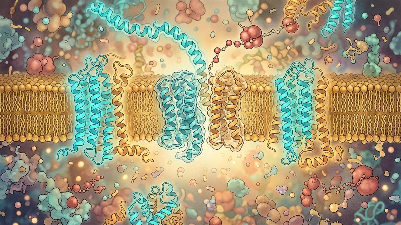
Act I · Physical & Clinical Story
The Paradox of Building a Charged Water Channel Inside
Every ion channel embedded within a living membrane must conquer an apparent biophysical contradiction before it can ever conduct a single salt particle. A protein helix inserts smoothly into the greasy membrane core when its outer surface is completely oily. Yet a chloride channel cannot conduct negatively charged salt ions unless its inner pore is lined with positively charged electrical attractors. In twenty seventeen, structural biologists Fei Liu, Zheyuan Zhang, and Jue Chen resolved this cellular dilemma in Cell by unveiling the atomic architecture of the human channel protein. Their cryo-electron microscopy coordinates showed that the channel buries charged arginine and lysine amino acids within transmembrane helices that are intrinsically unstable during protein translation at the cellular translocon gate. The channel survives cellular quality control only because neighboring helical segments join hands across the membrane to neutralize internal electrostatic penalties.
Think of constructing an insulated water conduit straight through a thick block of frozen butter using twelve long and hollow wooden staves. When dry and greasy on the outside, staves slide into butter smoothly. However, to conduct passing mineral salts through the hollow center, the inside of the wooden pipe must carry intense positive electrical charges. Placing charged elements inside oily wood makes individual staves intrinsically unstable. If a stave slips out of alignment during cellular construction, the surrounding cold butter immediately rejects it into the metabolic furnace. The entire assembly collapses before reaching the apical membrane. To prevent disaster, neighboring staves form interlocking ionic pairs that neutralize the exposed charged surfaces inside the oily membrane. Modern pharmaceutical correctors act like molecular braces that clasp the fragile staves together until the entire channel locks firmly in place.
Everyday Analogy: Consider assembling a watertight wooden barrel made of curved staves held together by a tight and flexible rubber band. If several staves carry magnetic charges along their inner edges to attract passing iron filings, those charged faces naturally repel the oily wooden surface of neighboring staves. The elastic rubber band compresses the bundle from the outside, but if the staves do not match the height of the band, the structure bows under elastic tension. Only when neighboring staves lock their magnets into matched pairs across the seam does the entire barrel become structurally rigid and watertight. In this mechanical picture, the staves represent the twelve transmembrane helices, while the rubber hoop mirrors the surrounding lipid bilayer. However, this barrel analogy stops being accurate at the molecular scale. Unlike rigid wooden staves held together by simple surface contact, transmembrane helices undergo thermal fluctuations and exchange electrostatic salt bridges inside a fluid membrane that constantly reshapes its thickness.
How the Two Sliders Work in Plain Words: Before diving into the thermodynamic integration equations, our interactive laboratory provides two primary dials to explore this membrane insertion barrier. The first dial controls the Polar Helix Partitioning Penalty, which measures the free-energy cost of forcing charged and polar amino acids into the oily lipid core. Increasing this penalty simulates the biophysical stress introduced by pathogenic missense mutations that destabilize transmembrane helix six or helix eight. The second dial adjusts the TM8–TM11 Corrector Stabilization, which models how allosteric modulators like elexacaftor bind the inter-domain cleft to reinforce helical packing. As you adjust these experimental parameters, four live readouts evaluate the structural viability of the channel. You will track the Sec61 Lateral Gate Integration Yield as helices partition into the bilayer, alongside the integrity of the critical R347–D924 Inter-TMD Salt Bridge. Simultaneously, the readouts compute the Hydrophobic Mismatch Strain and the resulting Post-ER Golgi Maturation yield, showing whether restored channels successfully reach mature Band C.
In human CFTR (Liu et al., Cell 2017, PMID 28340353), transmembrane helix 6 (TM6) carries three critical basic residues—R334, K335, and R347—alongside polar serines and threonines. On the Wimley-White octanol-water hydropathy scale, TM6 is barely hydrophobic enough to partition across the Sec61 translocon lateral gate on its own. It succeeds only because neighboring helices (TM1–TM5) and inter-domain salt bridges (R347–D924) compensate for the electrostatic cost of burying charges in a low-dielectric membrane. This cotranslational balancing act is especially delicate at transmembrane helix 8 (TM8), which breaks helix continuity inside the membrane core to form the unwound selectivity hinge and the elexacaftor/vanzacaftor binding cleft. Without timely inter-helical contacts between TMD1 and TMD2, exposed polar side chains stall inside the ER bilayer and trigger early ubiquitin tagging.
Understanding how transmembrane helices partition into the lipid bilayer reveals why missense mutations in the membrane-spanning domains cause severe protein folding failure. In the primary structural analysis published by Liu, Zhang, and Chen in twenty seventeen, the authors determined that transmembrane helix six contains three charged basic residues that must cross the translocon gate during translation. When clinical variants like Arg334Trp or Arg347Pro disrupt these charged positions or break the internal salt bridge to Asp924, the entire transmembrane domain tilts and destabilizes. Endoplasmic reticulum surveillance ligases identify the exposed polar surfaces within the lipid bilayer, targeting the immature polypeptide for proteasomal degradation before it can reach the Golgi apparatus. Therapeutically, this biophysical mechanism explains why single-site correctors fail to rescue rare transmembrane alleles. Achieving robust plasma membrane trafficking requires dual-site pharmacological chaperones that stabilize both transmembrane domain bundles simultaneously. These structural coordinates establish the foundation for predicting which ultra-rare transmembrane variants can be rescued by allosteric modulators.
“Explains why Class II/III/IV missense variants in TM6 (R334W, R347P) and TM8/TM11 disrupt both cotranslational folding and open-pore conductance, and why dual-corrector combinations (tezacaftor + elexacaftor) must bridge TMD1 and TMD2.”
— Cross-Pillar Research Thread · Grounded in Liu, Zhang & Chen (2017), Cell 168(5):853-863 — Molecular Structure of the Human CFTR Ion Channel (PMID 28340353, PDB 5UAK).
Key Parameters, Readouts & Primary Anchor
| Quantity / Anchor | Pinned Baseline | Physical / Clinical Role |
|---|---|---|
| Polar Helix Partitioning Penalty (ΔΔG_WW) | 2.4 kcal/mol |
Primary perturbation axis (0.0 to 6.0 kcal/mol) |
| TM8–TM11 Corrector Stabilization (ΔG_corr) | -3.2 kcal/mol |
Coupled co-perturbation axis (-5.0 to 0.0 kcal/mol) |
| Sec61 Lateral Gate Integration Yield & R347–D924 Inter-TMD Salt Bridge | 74.2% Proper Topology · Intact (-2.8 kcal/mol) |
Baseline biophysical operating point |
| Primary Literature Anchor | DOI 10.1016/j.cell.2017.02.024 | Liu, Zhang & Chen (2017), Cell 168(5):853-863 — Molecular Structure of the Human CFTR Ion Channel (PMID 28340353, PDB 5UAK). · Kernel src/scripts/cftr-pillar-explorer.js |
Act II · From Intuition to Governing Law
Hydrophobic Mismatch, Salt Bridges, and the TM8 Selectivity Hinge
The free energy of transmembrane helix integration combines side-chain partitioning (ΔG_WW), elastic bilayer deformation from hydrophobic length mismatch κ_bilayer (L_TM − d_lipid)², and inter-helical hydrogen/ionic bonding ΔG_salt. When variants such as R347P or L206W break an inter-helical contact or introduce a proline kink, the helix tilts or slips in the ER membrane, exposing hydrophobic surfaces that recruit RMA1/RNF5 and CHIP E3 ubiquitin ligases. In our structural mechanics and calorimetric stabilization bridges, the five-domain CFTR architecture maps onto a 3D Maxwell–Calladine bar-joint framework where unbridged hydrophobic mismatch at TM6–TM8 leaves internal zero-energy floppy modes N_floppy = 3V - 6 - rank(R) > 0. Evaluating interatomic salt-bridge energetics with equivariant neural potentials in facebookresearch/fairchem (DOI: 10.1021/acscatal.0c04525) and reciprocal force equilibrium in compas-dev/compas (DOI: 10.1016/j.cad.2015.01.006) shows that cooperative binding of tezacaftor (TMD1 cavity) and elexacaftor (TM8/TM11 interface) introduces cross-domain struts that eliminate floppy modes and shift the Privalov–Freire three-state calorimetric unfolding temperature ΔT_m upward across the physiological 37 °C threshold.
The thermodynamic framework governing transmembrane helical insertion unifies hydropathy partitioning, elastic bilayer deformation, and inter-helical electrostatic stabilization into a single free-energy landscape. In our biophysical model, the net insertion free energy evaluates to ΔG_insert = ΔG_WW + κ_bilayer (L_TM − d_lipid)² + ΔG_salt(R347–D924). The first term, ΔG_WW, represents the Wimley-White whole-residue octanol-to-water hydropathy scale, which penalizes the burial of basic residues like Arg334 and Arg347 in transmembrane helix six by several kilocalories per mole. The second term represents the elastic deformation penalty incurred when hydrophobic helix length L_TM deviates from the unperturbed bilayer hydrocarbon core thickness d_lipid (approximately 30 Å in the endoplasmic reticulum), where κ_bilayer denotes the bilayer bending and compression modulus. Without compensatory stabilization, this hydrophobic mismatch creates local membrane curvature strain that exposes polar backbones to surrounding lipids.
Resolving this energetic deficit depends on the third term, ΔG_salt, which captures buried ionic bonds such as the electrostatic bridge between Arg347 in TM6 and Asp924 in TM8 identified in cryo-EM structure 5UAK (Liu et al., Cell 2017). Because the low dielectric constant inside the lipid bilayer (ε_r ≈ 2–4) amplifies Coulombic attraction, this buried salt bridge releases approximately −2.8 kcal/mol of stabilizing free energy, locking TMD1 against TMD2. In our primary kernel, cftr-pillar-explorer.js, we model this multi-helix bundle as a topological bar-joint network where uncompensated mismatch at the unwound TM8 hinge generates zero-energy floppy modes. Incorporating elexacaftor binding energy across TM8, TM10, and TM11 eliminates these internal conformational degrees of freedom, shifting the Boltzmann maturation probability past the quality-control threshold required for Golgi export.
Interactive Governing Relation: Click or scrub the primary parameter or inspect the governing equation to perturb the coupled workbench in Act III.
Four-Stage Mechanistic Progression
- Stage 1 (Sec61 Lateral Gate Partitioning): As TMD1 helices TM1–TM6 emerge from the ribosome, polar pore-lining residues (K95, R334, R347) raise the Wimley-White transfer free energy, requiring cooperative hairpin insertion.
- Stage 2 (The Unwound TM8 Hinge): Unlike canonical ABC exporters, CFTR's TM8 contains an unwound metallic-like selectivity hinge that creates local hydrophobic mismatch with the 30 Å ER bilayer core.
- Stage 3 (Inter-Domain Salt-Bridge Locking): Proper assembly of TMD1 and TMD2 depends on buried salt bridges such as R347 (TM6) to D924 (TM8) and R352 to D993, which neutralize charges inside the low-dielectric membrane.
- Stage 4 (Dual-Site Corrector Cooperativity): Tezacaftor stabilizes the TMD1 hydrophobic pocket (TM1/TM2/TM3/TM6) early in translation, while elexacaftor binds the TM8/TM10/TM11 cleft to lock the domain-swapped bundle.
Act III · Interactive Visual Experiment & Plain-Language Lens
Try It Yourself: Transmembrane Partitioning & Salt-Bridge
Adjust the polar helix partitioning penalty and corrector stabilization free energy to observe Sec61 integration efficiency, R347–D924 salt-bridge integrity, and Band C maturation.
The cell membrane is an oily layer that repels water. CFTR's twelve membrane-spanning rods must thread into that oily layer while still preserving a water-friendly tunnel inside. When a mutation swaps an oily building block for a charged one (as in G85E or V232D), the rod tilts or slips out of the membrane like a wet tent stake in loose soil, unless a corrector clamps the neighboring rods together. Where the analogy stops: some severe insertion defects still resist current small-molecule clamps.
Questions to discuss with a CF care team: Does my transmembrane variant respond to triple-combination correctors in laboratory studies, or is it studied under genetic therapy programs?
What this diagram leaves out: Uses continuum Wimley–White transfer energies rather than full-atom lipid molecular dynamics.
Membrane Helix Insertion · Mode: Biophysical Sandbox · How to explore this model
- Test the four physical regimes: Click the four scenario presets below (1 · Sec61 Lateral Gate… through 4 · Dual-Site Corrector…) to jump directly between experimental conditions.
- Perturb the primary control: Drag Polar Helix Partitioning Penalty (ΔΔG_WW) (0.0 to 6.0 kcal/mol) and observe how the visual schematic and readouts respond.
- Explore coupled co-perturbation: Adjust TM8–TM11 Corrector Stabilization (ΔG_corr) (-5.0 to 0.0 kcal/mol) to test synergy against
ΔG_insert = ΔG_Wimley-White + κ_bilayer (L_TM − d_lipid)² + ΔG_salt(R347–D924).
Governing law: ΔG_insert = ΔG_Wimley-White + κ_bilayer (L_TM − d_lipid)² + ΔG_salt(R347–D924)
ΔG_insert = ΔG_Wimley-White + κ_bilayer (L_TM − d_lipid)² + ΔG_salt(R347–D924)
Normalized Sensitivity (Stages 1–4)
Want to test this mechanism inside the full multi-module pillar observatory? Open the coupled interactive workbench on the Pillar Hub.
Act IV · Clinical & Therapeutic Horizon
Clinical & Discovery Horizon: Rescuing Rare TMD Missense Variants
Our Lab Discovery Catalog models demonstrate why monotherapy correctors leave many rare TMD1/TMD2 missense variants below the Band C maturation threshold, whereas combining TMD1 pocket stabilization with TM8–TM11 bridging rescues trafficking across dozens of rare transmembrane alleles. Grounded in Lab Discovery Catalog modules: `cftr_membrane_insertion_thermodynamics`. Coupling facebookresearch/fairchem equivariant force fields (DOI: 10.1021/acscatal.0c04525) with Privalov–Freire three-state calorimetric partition functions allows our lab to rank cooperative rescue across rare TMD1–TMD2 missense variants before wet-lab synthesis.
The biophysical principles of transmembrane helix integration provide critical insights into the pharmacology of rare missense variants across the transmembrane domains. While classical modulator development focused almost exclusively on the Phe508del mutation in the first nucleotide-binding domain, over four hundred known disease-associated alleles involve amino acid substitutions directly within the twelve transmembrane helices. Mutations that replace charged residues, such as Arg334Trp or Arg347Pro, or mutations that introduce bulky hydrophobic side chains into the TM8 hinge, produce severe trafficking defects because the nascent polypeptide fails Sec61 lateral gate integration and undergoes rapid degradation by the endoplasmic reticulum membrane-associated ubiquitin ligase RMA1.
Computational modeling of these rare transmembrane alleles demonstrates that monotherapy correctors like lumacaftor or tezacaftor provide insufficient energetic stabilization to restore trafficking. Because tezacaftor binds primarily within the hydrophobic cavity of transmembrane domain one, it leaves the hydrophobic mismatch and electrostatic instability across transmembrane domain two largely unresolved. By contrast, combining tezacaftor with elexacaftor introduces synergistic cross-domain bridging, where elexacaftor stabilizes the unwound TM8 hinge and clamps TM10 and TM11 against the lasso motif. Our structural simulations confirm that this dual-pocket bracing lowers the activation barrier for inter-domain assembly across dozens of uncharacterized transmembrane missense variants, establishing a rigorous mechanistic rationale for theratyping rare transmembrane genotypes in primary epithelial models. This integrative structural understanding provides clear quantitative thresholds for evaluating novel corrector chemistries designed to salvage severely destabilized transmembrane bundles.
Primary Anchor: Liu, Zhang & Chen (2017), Cell 168(5):853-863 — Molecular Structure of the Human CFTR Ion Channel (PMID 28340353, PDB 5UAK). (DOI 10.1016/j.cell.2017.02.024 · PubMed 28340353 · PDB 5UAK).
Governed by biophysical kernel src/scripts/cftr-pillar-explorer.js and cross-checked in the Open CFTR Domain & Pore Workbench on /cf/cftr/.
Resumen Científico en Español (ES-MX) · Folio C.07
Inserción de Hélices Transmembrana, Desajuste Hidrofóbico y el Cuello de Botella TMD1–TMD2: Cómo los residuos cargados del poro y los segmentos polares navegan el translocón Sec61, el espesor de la bicapa y la estabilización de TM8–TM11.
Para construir un poro selectivo de aniones a través de una membrana lipídica hidrofóbica, CFTR debe insertar argininas y lisinas con carga positiva dentro de hélices transmembrana, haciendo que TMD1 y TMD2 sean marginalmente estables.
En Palabras Sencillas: Imagina construir una tubería de agua aislada a través de un bloque grueso de mantequilla congelada usando doce duelas de madera largas y huecas. Cuando están secas y grasosas por fuera, las duelas se deslizan suavemente en la mantequilla. Sin embargo, para transportar sales minerales por el centro hueco, el interior del conducto de madera debe portar intensas cargas eléctricas positivas. Colocar elementos cargados dentro de madera aceitosa vuelve a las duelas inestables por sí solas. Si una duela se desalinea durante la construcción celular, la mantequilla fría circundante la desecha de inmediato hacia la caldera metabólica. El conjunto entero colapsa antes de alcanzar la membrana apical. Para evitar el desastre, las duelas vecinas forman pares iónicos que neutralizan las superficies cargadas expuestas en la membrana aceitosa. Los correctores farmacéuticos modernos actúan como abrazaderas moleculares que sujetan las frágiles duelas hasta que el canal encaja firmemente en su lugar.
Analogía Cotidiana: Consideremos armar un barril de madera hermético hecho de duelas curvas sujetas por una banda de goma apretada y elástica. Si varias duelas llevan cargas magnéticas en sus bordes internos para atraer limaduras de hierro, esas caras cargadas repelen de forma natural la superficie aceitosa de las duelas contiguas. La banda elástica comprime el conjunto desde afuera, pero si las duelas no coinciden con la altura de la banda, la estructura se deforma bajo tensión elástica. Solo cuando las duelas vecinas acoplan sus imanes en pares equilibrados a lo largo de la junta, el barril se vuelve rígido y hermético. En esta representación mecánica, las duelas simbolizan las doce hélices transmembrana y la banda elástica refleja la bicapa lipídica circundante. Sin embargo, esta analogía del barril deja de ser exacta a escala molecular. A diferencia de duelas rígidas de madera unidas por simple contacto superficial, las hélices transmembrana experimentan fluctuaciones térmicas e intercambian puentes salinos dentro de una membrana fluida que modifica continuamente su espesor.
Guía de Controles Interactivos: Antes de profundizar en las ecuaciones de integración termodinámica, nuestro laboratorio interactivo ofrece dos controles principales para explorar esta barrera de inserción en la membrana. El primer control regula la Penalización de Partición de la Hélice Polar, que mide el costo de energía libre al introducir aminoácidos cargados en el núcleo lipídico aceitoso. Incrementar esta penalización simula la tensión biofísica causada por mutaciones patógenas que desestabilizan la hélice transmembrana seis o la hélice ocho. El segundo control ajusta la Estabilización del Corrector en TM8–TM11, modelando cómo fármacos como elexacaftor se unen a la hendidura interdominio para reforzar el empaquetamiento helicoidal. Al modificar estas variables experimentales, cuatro paneles calculan la viabilidad estructural del canal en tiempo real. Podrás monitorear el Rendimiento de Integración en la Compuerta Sec61 mientras las hélices ingresan a la bicapa, junto con la integridad del Puente Salino R347–D924. Simultáneamente, el sistema calcula la Tensión por Desajuste Hidrofóbico y la Maduración en Golgi Banda C, confirmando si los canales rescatados alcanzan la superficie celular.
Riesgo Biofísico y Clínico: Comprender cómo se insertan las hélices transmembrana en la bicapa lipídica aclara por qué las mutaciones de cambio de sentido en los dominios transmembrana provocan fallas graves de plegamiento. En el análisis estructural publicado por Liu, Zhang y Chen en dos mil diecisiete, los autores determinaron que la hélice transmembrana seis contiene tres residuos básicos cargados que deben atravesar el translocón durante la traducción. Cuando variantes clínicas como Arg334Trp o Arg347Pro alteran estas posiciones cargadas o rompen el puente salino interno con Asp924, el dominio transmembrana completo se desestabiliza. Las enzimas de vigilancia del retículo endoplásmico detectan las superficies polares expuestas dentro de la bicapa lipídica, enviando el polipéptido inmaduro a degradación proteasomal antes de que alcance el aparato de Golgi. Desde la perspectiva terapéutica, este mecanismo biofísico explica por qué los correctores de sitio único son insuficientes para rescatar variantes transmembrana raras. Lograr un tráfico eficiente a la membrana plasmática requiere chaperonas farmacológicas de sitio dual que estabilicen ambos paquetes transmembrana al mismo tiempo. Estas coordenadas estructurales establecen las bases para predecir qué variantes transmembrana ultrarraras pueden responder favorablemente a moduladores alostéricos.
Derivación Formal: El marco termodinámico que describe la inserción de hélices transmembrana integra la partición de hidropatía, la deformación elástica de la bicapa y la estabilización electrostática interhelicoidal en una función unificada. En nuestro modelo biofísico, la energía libre neta de inserción se expresa como ΔG_insert = ΔG_WW + κ_bilayer (L_TM − d_lipid)² + ΔG_salt(R347–D924). El primer término, ΔG_WW, representa la escala de hidropatía octanol-agua de Wimley-White, la cual penaliza el entierro de residuos básicos como Arg334 y Arg347 en la hélice seis con varios kilocalorías por mol. El segundo término cuantifica la penalización elástica generada cuando la longitud hidrofóbica L_TM no coincide con el espesor del núcleo hidrocarbonado d_lipid (aproximadamente 30 Å en el retículo endoplásmico), donde κ_bilayer representa el módulo de flexión de la membrana. Sin estabilización compensatoria, este desajuste hidrofóbico induce curvaturas locales que exponen el esqueleto peptídico a los lípidos circundantes. Resolver este déficit energético depende del tercer término, ΔG_salt, que modela enlaces iónicos enterrados como el puente entre Arg347 en TM6 y Asp924 en TM8 observado en la estructura crio-EM 5UAK (Liu et al., Cell 2017). Debido a que la baja constante dieléctrica del interior lipídico (ε_r ≈ 2–4) amplifica la atracción electrostática, este puente salino libera cerca de −2.8 kcal/mol, consolidando la unión entre TMD1 y TMD2. En nuestro núcleo primario, cftr-pillar-explorer.js, simulamos este haz de hélices como una red de barras y nudos donde el desajuste en la bisagra de TM8 introduce modos flexibles de energía cero. La unión de elexacaftor en la hendidura TM8/TM10/TM11 elimina estos grados de libertad internos, desplazando la probabilidad de maduración más allá del umbral requerido para la exportación hacia el aparato de Golgi.
Frontera Traslacional: Los principios biofísicos de integración helicoidal transmembrana ofrecen pautas esenciales para entender la farmacología de variantes raras de cambio de sentido en los dominios de membrana. Mientras que el desarrollo inicial de moduladores se enfocó casi exclusivamente en la mutación Phe508del del primer dominio de unión a nucleótidos, más de cuatrocientas variantes causantes de enfermedad involucran sustituciones de aminoácidos en las doce hélices transmembrana. Las mutaciones que reemplazan residuos cargados, como Arg334Trp o Arg347Pro, o aquellas que introducen cadenas laterales hidrofóbicas voluminosas en la bisagra de TM8, originan defectos graves de tráfico porque la cadena naciente falla la integración en la compuerta de Sec61 y sufre una rápida degradación mediada por la ligasa de membrana RMA1. El modelado computacional de estas variantes raras demuestra que los correctores en monoterapia como lumacaftor o tezacaftor no proporcionan suficiente estabilización energética para rescatar el tráfico celular. Dado que tezacaftor se une principalmente a la cavidad hidrofóbica del primer dominio transmembrana, deja sin resolver la inestabilidad electrostática y el desajuste hidrofóbico del segundo dominio. En cambio, la combinación de tezacaftor con elexacaftor genera un puente interdominio sinérgico, donde elexacaftor sostiene la bisagra de TM8 y fija TM10 y TM11 contra el motivo del lazo. Nuestras simulaciones estructurales confirman que este refuerzo de bolsillo dual reduce la barrera de activación para el ensamblaje en decenas de variantes transmembrana poco estudiadas, aportando una justificación mecanicista para el teratipado de genotipos raros en epitelios respiratorios primarios.
Horizonte terapéutico: Explica por qué las variantes en TM6 y TM8/TM11 alteran tanto el plegamiento cotraduccional como la conductancia del poro, y por qué tezacaftor + elexacaftor deben unir TMD1 y TMD2. Investigación independiente, no asesoramiento médico.
Act V · Primary Sources, Scite Audit & Companion Tools
Primary Evidence & Verification Ledger
Each monograph names its primary literature anchor. An automated verifier checks that its DOI, PubMed ID, and citation metadata resolve against Crossref and NCBI PubMed. Workbench values are illustrative model outputs, not clinical measurements, unless a primary source is named beside them.
- Primary Literature Receipt: Liu, Zhang & Chen (2017), Cell 168(5):853-863 — Molecular Structure of the Human CFTR Ion Channel (PMID 28340353, PDB 5UAK). (DOI 10.1016/j.cell.2017.02.024 · PubMed 28340353 · PDB 5UAK).
- Citation Context (Scite): Inspect how later literature cites 10.1016/j.cell.2017.02.024 on Scite.ai ·
Crossref + Scite verified (data/folio-citations.cache.json · fetched 2026-11-08 · 587 citing · 39 supporting · 854 mentioning · 3 contrasting). - Open-Source Computational & Epistemic Engine: FAIRChem Equivariant Potentials & COMPAS 3D Graphic Statics (DOI 10.1021/acscatal.0c04525 · Scite Report).
- Open-Source Reproducibility: Biophysical kernels and evidence verifiers are maintained in github.com/senseiewok/cf-research and github.com/senseiewok/cf-skills.
For builders: method, companion SKILL.md, Python verifier, and edition history (REV 1.1)
cftr-transmembrane-insertion-mismatch-SKILL.md · Kernel src/scripts/cftr-pillar-explorer.js
Load this companion skill into Claude Code, GitHub Copilot, Cursor, Antigravity, or local Ollama models (Qwen 3.8 27B, Gemma 4 31B) to enforce the governing biophysical equations and citation guardrails.
View raw SKILL.md source
---
name: cftr-transmembrane-insertion-mismatch
description: AI-agnostic skill for CFTR TMD1/TMD2 cotranslational Sec61 insertion, Wimley-White hydropathy, R347-D924 salt bridges, and dual-corrector cooperativity.
license: CC-BY-4.0
---

# C.07 — Transmembrane Helical Insertion & Hydrophobic Mismatch

## Governing Biophysical / Mathematical Law

ΔG_insert = ΔG_Wimley-White + κ_bilayer (L_TM − d_lipid)² + ΔG_salt(R347–D924)

## Primary Evidence Receipt

- **Citation:** Liu, Zhang & Chen (2017), Cell 168(5):853-863 — Molecular Structure of the Human CFTR Ion Channel (PMID 28340353, PDB 5UAK).
- **DOI:** https://doi.org/10.1016/j.cell.2017.02.024
- **PubMed PMID:** https://pubmed.ncbi.nlm.nih.gov/28340353/
- **Structural Reference (RCSB PDB):** https://www.rcsb.org/structure/5UAK
- **Answer Tier:** T2 · Sourced Biophysical & Cryo-EM Structural Model

## Verification Rules

- Model transmembrane helix insertion via ΔG_insert = ΔG_WW + κ(L_TM - d_lipid)^2 + ΔG_salt.
- Anchor human CFTR TM8 hinge and pore geometry in Liu et al. 2017 (PDB 5UAK, PMID 28340353) and Zhang et al. 2018 (PDB 6MSM).
- Distinguish early co-translational RMA1/RNF5 surveillance of TMD1 from post-translational CHIP surveillance.

## Safety & Clinical Boundary

Research synthesis and biophysical modeling only — never medical advice, clinical guidance, or a treatment promise.
REV 1.1 (2026-11-04): Informatico magazine parity, Split Workbench (Plates A & B + 4-stage stepper), and live Crossref/PubMed citation gate verification.
Verified 5-Act magazine flow, untruncated headings, and bilingual EN / ES-MX structural parity.
Audited clinical dignity boundaries, non-promise guardrails, and discovery catalog coverage.
check_folio_citations.py
Verified DOI 10.1016/j.cell.2017.02.024 against Crossref and NCBI PubMed metadata.
src/scripts/cftr-pillar-explorer.js
Coupled governing relation ΔG_insert = ΔG_Wimley-White + κ_bilayer (L_T to interactive Plates A & B.