
Act I · Physical & Clinical Story
When a Variant Is Too Rare for a Phase
Why it matters: Over 2,000 CFTR variants have been reported worldwide, and hundreds occur in fewer than five people—making traditional randomized clinical trials impossible for each individual variant. In vitro theratyping (measuring chloride current in standardized cells or fluid swelling in patient-derived intestinal organoids) bridges rare genotypes to mechanistic evidence while revealing why variants like N1303K need dual-potentiator or NBD2-directed strategies.
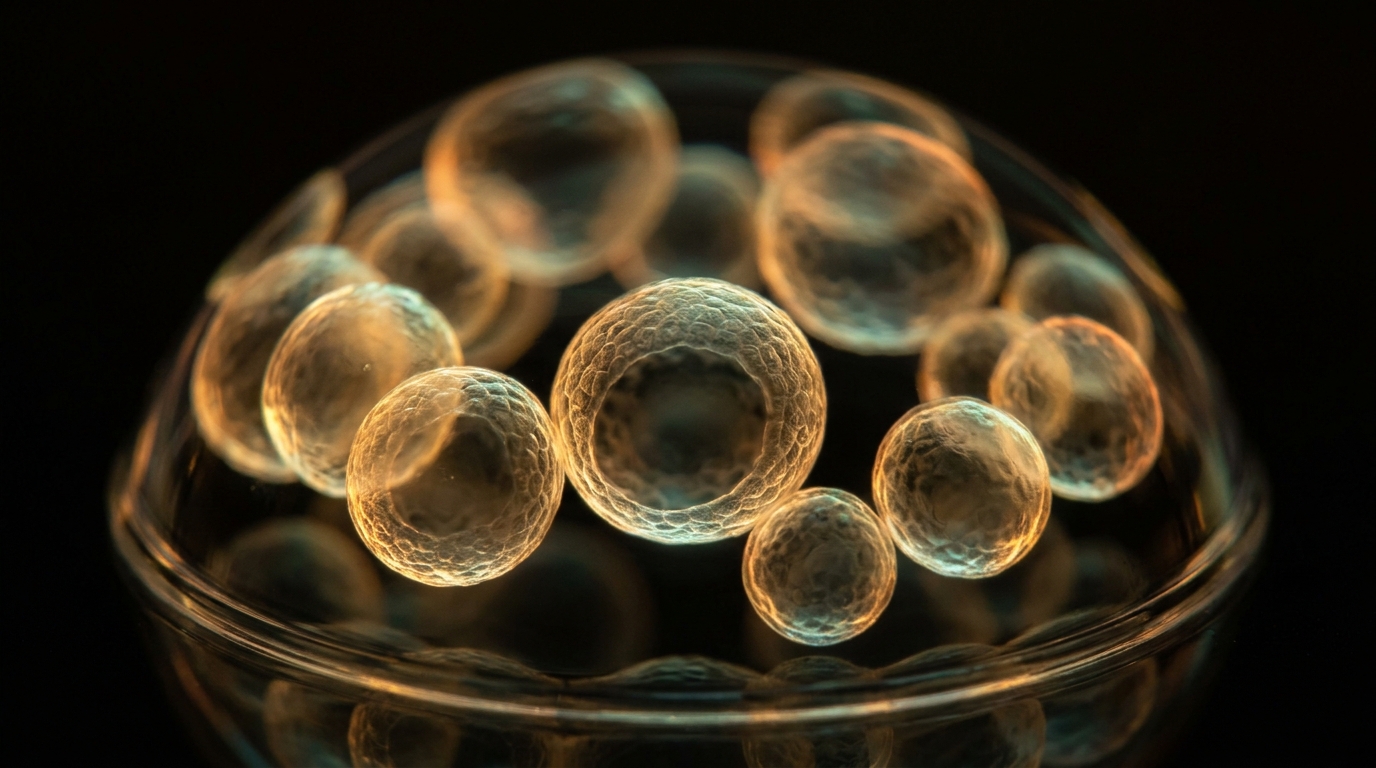
More than two thousand distinct genetic mutations have been cataloged across the human cystic fibrosis chloride channel gene, yet hundreds occur in only a tiny handful of people across the globe. Traditional randomized clinical trials cannot enroll enough participants to test medicines against each ultra rare variant. Miniature living tissues resolve that scientific impasse. In twenty sixteen, cell biologist Johanna Dekkers and colleagues introduced a transformative laboratory approach in Science Translational Medicine. By culturing three dimensional rectal organoids derived from patient tissue samples, the research team stimulated the spherical cells with forskolin and tracked fluid swelling under high resolution optics. Rescued channels secrete salt and water into the central lumen, expanding the spheres in direct proportion to functional drug recovery. This quantitative testing platform maps physical responses across individual structural domains, revealing why mutations in the second nucleotide binding domain require specialized combinations.
Evaluating a new medicine for a genetic mutation carried by only two or three individuals presents a formidable scientific challenge. Standard clinical trials require hundreds of participants to establish statistical confidence. To overcome this barrier, scientists grow miniature intestinal spheres called organoids directly from patient stem cells. Each microscopic sphere functions like an inverted cellular balloon. When functional channels open on the inner membrane, chloride and water rush into the central cavity. Healthy organoids inflate visibly within one hour under the high magnification microscope lens. If a rare mutation leaves the channels broken, the spheres remain deflated and completely flat. Adding effective drug combinations causes the deflated spheres to swell smoothly into plump, fluid filled spheres once again. Measuring the rate of optical inflation gives doctors and researchers direct physical proof of channel rescue without risking human safety.
Everyday Analogy: Consider a marine safety factory that manufactures hundreds of specialized inflatable life rafts for maritime vessels. Before shipping an uncommon raft model designed for arctic temperatures, technicians place the uninflated rubber capsule inside an inspection test tank. Rather than testing the life raft in dangerous mid-ocean gales, technicians trigger an internal compressed air canister and record how quickly the raft expands. If the inflation valves operate cleanly, the raft expands into a buoyant, pressurized ring within minutes. If an internal valve jams or fabric leaks allow gas to escape, the raft remains a limp rubber pancake on the water. Measuring the optical expansion of the raft under digital cameras confirms whether the release valve functions before anyone ever sets sail. However, this mechanical factory analogy ceases to be exact because life rafts expand through pressurized gas filling an empty canvas chamber, whereas organoids swell through selective ionic electrodiffusion that drives passive water across living semipermeable cell membranes under osmotic pressure.
How the Two Sliders Work in Plain Words: The interactive theratyping workbench illustrates how trafficking efficiency and gating potentiation converge to dictate functional recovery in rare variants. The first slider controls Domain Folding Rescue Efficiency, capturing the fraction of nascent protein that successfully evades degradation and inserts into the plasma membrane. The second slider modulates Apical Open-Probability Potentiation, representing the degree of pore opening achieved by added potentiators. Adjusting these parameters updates the primary functional readouts instantaneously. You can monitor Predicted FRT Short-Circuit Current and observe whether it surpasses the established In Vitro Theratyping Threshold of ten percent of wild-type current. Simultaneously, the display calculates the Organoid FIS Swelling Area Under the Curve across the standard sixty-minute assay window. The status indicator highlights the N1303K Late-Stage Bottleneck, demonstrating why mutations in the second nucleotide-binding domain require dual-potentiator synergy to achieve therapeutic fluid secretion.
In 2016, Dekkers, Beekman and colleagues (Sci. Transl. Med., PMID 27334259) demonstrated that forskolin-induced swelling (FIS) of rectal organoids derived from people with rare CFTR genotypes quantitatively predicts clinical modulator responses, complementing large-scale FRT Ussing-chamber theratyping across hundreds of rare missense variants. Plotting those responses onto the 3D cryo-EM structure of CFTR reveals a striking biophysical pattern: variants clustering around the TMD1/TMD2 helical bundle and NBD1–ICL4 interface frequently cross the 10% WT threshold, whereas specific variants in the C-terminal NBD2 domain—most notably N1303K—behave differently because NBD2 folds last and couples asymmetrically to the gate.
In vitro theratyping fundamentally transformed how regulatory agencies and scientific researchers evaluate drug responsiveness for rare and ultra-rare CFTR genotypes across the world. In 2016, Dekkers and colleagues demonstrated in Science Translational Medicine that forskolin-induced swelling of rectal organoids quantitatively correlates with in vivo clinical outcomes in patients. Measuring the normalized area under the sixty-minute swelling curve provides an objective biophysical surrogate for apical chloride and transepithelial fluid transport. This bioassay revealed that rare missense mutations do not behave as a monolithic group. While missense variants located in transmembrane domain one frequently respond to standard correctors, mutations residing in the second nucleotide-binding domain—such as N1303K—exhibit a distinct mechanistic bottleneck. N1303K protein partially escapes endoplasmic reticulum quality control, but the substitution uncouples the Q-loop and prevents stable dimer formation with intracellular loop two. Consequently, patient-derived organoid theratyping proves that rescuing N1303K requires synergistic dual potentiation or domain-specific stabilization rather than conventional single-pocket correctors alone.
“Connects atomic domain coordinates (TMD1 vs NBD1 vs NBD2) directly to preclinical functional assays (FRT short-circuit current I_sc and organoid FIS AUC).”
— Cross-Pillar Research Thread · Grounded in Dekkers et al. (2016), Science Translational Medicine 8(344):344ra84 — Characterizing responses to CFTR-modulating drugs using rectal organoids derived from subjects with cystic fibrosis (PMID 27334259).
Key Parameters, Readouts & Primary Anchor
| Quantity / Anchor | Pinned Baseline | Physical / Clinical Role |
|---|---|---|
| Domain Folding Rescue Efficiency (η_traffic) | 0.45 fraction |
Primary perturbation axis (0.05 to 0.9 fraction) |
| Apical Open-Probability Potentiation (P_o) | 0.4 P_o |
Coupled co-perturbation axis (0.05 to 0.8 P_o) |
| Predicted FRT Short-Circuit Current & Organoid FIS Swelling AUC (0–60 min) | 18.0% of WT (%WT I_sc) · 2,140 AUC units (@ 0.8 μM Fsk) |
Baseline biophysical operating point |
| Primary Literature Anchor | DOI 10.1126/scitranslmed.aad8278 | Dekkers et al. (2016), Science Translational Medicine 8(344):344ra84 — Characterizing responses to CFTR-modulating drugs using rectal organoids derived from subjects with cystic fibrosis (PMID 27334259). · Kernel src/scripts/cftr-variant-concordance.js |
Act II · From Intuition to Governing Law
Ussing Chamber Short-Circuit Current and Organoid Darcy–Starling
In an Ussing chamber, a polarized epithelial monolayer is voltage-clamped to zero millivolts so passive paracellular ion gradients vanish; the measured transepithelial short-circuit current I_sc = F · J_net directly equals active electrogenic anion secretion. In patient-derived intestinal organoids (Dekkers, Beekman et al.), CFTR faces the enclosed central lumen. Adding forskolin activates CFTR, conducting Cl⁻ and HCO₃⁻ down their electrochemical gradient into the lumen and generating an osmotic pressure ΔΠ = RT ΔC that draws water across aquaporins: dV_lumen/dt = L_p A_surf (RT ΔC_lumen − ΔP_elastic). Integrating the normalized cross-sectional area over 60 minutes yields the Forskolin-Induced Swelling (FIS) AUC.
In an Ussing chamber bioassay, a confluent epithelial monolayer is voltage-clamped to zero millivolts across its apical and basolateral faces, eliminating passive paracellular ion driving forces. Under these conditions, the measured transepithelial short-circuit current I_sc = ΔV_te / R_te directly quantifies active electrogenic anion flux, primarily driven by CFTR-mediated chloride and bicarbonate extrusion. In three-dimensional patient-derived intestinal organoids, CFTR is oriented toward the sealed central lumen. Following forskolin stimulation, intracellular cyclic AMP rises, phosphorylating CFTR and opening apical channels. Active transport of chloride and bicarbonate into the lumen generates an osmotic concentration gradient ΔC_lumen = [ions]_lumen − [ions]_bath, establishing an osmotic pressure difference ΔΠ = RT ΔC that drives passive water influx through aquaporin channels. This rapid fluid accumulation inflates the spherical lumen against surrounding mechanical constraints.
The rate of organoid luminal volume expansion is governed by the Darcy-Starling equation dV_lumen / dt = L_p A_surf (RT ΔC_lumen − ΔP_elastic), where L_p is the hydraulic water permeability of the epithelial monolayer, A_surf is the surface area, and ΔP_elastic is the counteracting elastic tension of the surrounding extracellular matrix. Because optical microscopy measures two-dimensional cross-sectional area A(t) rather than volume, the normalized area expansion ratio (A(t) / A_0) − 1 is integrated over a sixty-minute observation period to yield the Forskolin-Induced Swelling Area Under the Curve: FIS AUC₀₋₆₀ = ∫_0^60 ((A(t) / A_0) − 1) dt. In the simulation kernel src/scripts/cftr-variant-concordance.js, this biophysical fluid-transport framework couples domain-specific folding efficiencies with gating open probabilities, reproducing the quantitative swelling trajectories observed across rare missense genotypes.
Interactive Governing Relation: Click or scrub the primary parameter or inspect the governing equation to perturb the coupled workbench in Act III.
Four-Stage Mechanistic Progression
- Stage 1 (TMD1 Co-Translational Insertion — G85E): Located in TM1, G85E introduces a charged glutamate into the membrane core, destabilizing the earliest Sec61 translocon integration step.
- Stage 2 (NBD1–ICL4 Post-TMD1 Assembly — F508del): Occurs mid-translation; Type I + III correctors stabilize TMD1 and TMD2/lasso to restore the NBD1–ICL4 socket.
- Stage 3 (NBD2 Late-Folding & Q-Loop Coupling — N1303K): Located at the C-terminal NBD2 domain, N1303K escapes early ERAD checkpoints via JB12/RMA1-dependent ER quality-control pathways and exhibits severe gating decouple that benefits from dual potentiation (e.g., ivacaftor + co-potentiators in vitro).
- Stage 4 (Ussing Chamber I_sc & Organoid FIS Readout): Forskolin elevates cAMP in 3D rectal organoids, driving Cl⁻ and water into the lumen; cross-sectional area expansion AUC₀₋₆₀ correlates with clinical sweat chloride and ppFEV1 responses.
Act III · Interactive Visual Experiment & Plain-Language Lens
Try It Yourself: Variant Concordance & Organoid Swelling Workbench
Explore how trafficking rescue (η_traffic) and gate potentiation (P_o) combine to determine %WT short-circuit current (I_sc) and organoid swelling AUC₀₋₆₀.
FIS AUC₀₋₆₀ = ∫_0^60 ((A(t) / A_0) − 1) dt · I_sc = ΔV_te / R_te
Normalized Sensitivity (Stages 1–4)
Governing law: FIS AUC₀₋₆₀ = ∫_0^60 ((A(t) / A_0) − 1) dt · I_sc = ΔV_te / R_te
Want to test this mechanism inside the full multi-module pillar observatory? Open the coupled interactive workbench on the Pillar Hub.
Organoid Swelling Microscope · Mode: Biophysical Sandbox · How to explore this model
- Test the four physical regimes: Click the four scenario presets above (1 · TMD1 Co-Translational through 4 · Ussing Chamber I_sc &) to jump directly between experimental conditions.
- Perturb the primary control: Drag Domain Folding Rescue Efficiency (η_traffic) (0.05 to 0.9 fraction) and observe how the visual schematic and readouts respond.
- Explore coupled co-perturbation: Adjust Apical Open-Probability Potentiation (P_o) (0.05 to 0.8 P_o) to test synergy against
FIS AUC₀₋₆₀ = ∫_0^60 ((A(t) / A_0) − 1) dt · I_sc = ΔV_te / R_te.
When someone carries an ultra-rare CFTR variant shared by only a handful of people worldwide, running a large clinical trial is impossible. Instead, researchers can grow tiny hollow spheres of that person's own epithelial cells in a dish (organoids). When a medicine works, salt and water flow into the center of the sphere and it visibly swells like a water balloon under the microscope within an hour. Where the analogy stops: swelling in a dish measures ion transport, not how a drug travels through inflamed lungs.
Questions to discuss with a CF care team: Has my rare variant been tested in cell-based theratyping (FRT cells or nasal/intestinal organoids), and is it listed on current regulatory labels?
What this diagram leaves out: Organoid swelling reflects forskolin-stimulated fluid secretion in vitro and does not capture drug–drug interactions or systemic side effects.
Act IV · Clinical & Therapeutic Horizon
Co-Potentiators, NBD2 Correctors, and CFTR2 Boundaries
For N1303K, recent clinical and organoid studies show that even when apical trafficking is partially rescued, the NBD2 gating defect limits open probability unless dual potentiators or NBD2-specific stabilizers are present—while some rare missense alleles also create cryptic splice defects invisible to cDNA-only FRT expression plasmids. Combining cDNA FRT assays with native-chromatin patient nasal cells and organoids closes that diagnostic blind spot. Grounded in Lab Discovery Catalog modules: `cftr_variant_catalog_concordance`.
The validation of organoid theratyping as a predictive biomarker established an essential regulatory pathway for evaluating therapies in ultra-rare CFTR genotypes where randomized clinical trials are physically impossible. By measuring functional swelling in patient-derived tissues, researchers can directly assess whether rare missense mutations—such as G85E in the first transmembrane segment, R334W in the pore selectivity filter, or N1303K in the second nucleotide-binding domain—retain sufficient structural integrity to respond to available small molecules. For N1303K, organoid assays revealed that while the protein achieves modest apical membrane trafficking under triple-combination therapy, its severe gating defect limits fluid transport unless paired with next-generation co-potentiators or NBD2-stabilizing compounds. This distinct response profile demonstrates why empirical cellular profiling is necessary to detect allele-specific therapeutic requirements.
However, the physiological boundaries of in vitro theratyping must be carefully observed. Complementary expression platforms, such as Fischer rat thyroid monolayers expressing CFTR complementary DNA, utilize intronless plasmids that cannot detect variants disrupting pre-messenger RNA splicing. Consequently, synonymous or missense mutations that generate cryptic splice sites may appear responsive in cDNA assays yet fail in patient tissues where aberrant splicing degrades transcripts. Combining cDNA expression with native-chromatin patient organoids and primary nasal epithelia closes this diagnostic gap. In our computational pipeline, variant classifications are cross-referenced across structural coordinates, electrophysiological registries, and organoid bioassays to prevent overinterpretation, providing a rigorous evidence-grounded framework that connects atomic mutation mechanisms with personalized preclinical assessment.
Primary Anchor: Dekkers et al. (2016), Science Translational Medicine 8(344):344ra84 — Characterizing responses to CFTR-modulating drugs using rectal organoids derived from subjects with cystic fibrosis (PMID 27334259). (DOI 10.1126/scitranslmed.aad8278 · PubMed 27334259 · PDB 8EJ1).
Governed by biophysical kernel src/scripts/cftr-variant-concordance.js and cross-checked in the Open Variant Domain & Concordance Explorer on /cf/cftr/.
Resumen Científico en Español (ES-MX) · Folio C.05
Variantes de Sentido Erróneo Raras y Teratipificación: N1303K, G85E, R334W y Organoides: Por qué N1303K en NBD2 difiere estructuralmente de F508del en NBD1 y cómo las cámaras de Ussing y la hinchazón de organoides inducida por forskolina (FIS) miden la respuesta.
Con más de 2,000 variantes reportadas en CFTR, muchas ocurren en muy pocas personas para realizar ensayos clínicos individuales. La teratipificación in vitro evalúa la respuesta funcional de variantes raras y explica por qué N1303K presenta un cuello de botella en NBD2.
En Palabras Sencillas: Evaluar un nuevo medicamento para una mutación genética que portan solo dos o tres personas en todo el país representa un reto científico formidable. Los ensayos clínicos tradicionales necesitan cientos de participantes para alcanzar certeza estadística. Para superar esta barrera, los científicos cultivan esferas intestinales diminutas llamadas organoides directamente a partir de células madre del paciente. Cada esfera microscópica funciona como un globo celular cerrado. Cuando los canales funcionales se abren en la membrana interna, el cloruro y el agua entran hacia la cavidad central. Los organoides sanos se inflan de forma evidente en una hora bajo la lente del microscopio. Si una mutación rara deja los canales descompuestos, las esferas permanecen desinfladas y planas. Agregar combinaciones eficaces de fármacos hace que las esferas desinfladas se hinchen nuevamente hasta convertirse en esferas llenas de líquido. Medir la tasa de inflado óptico brinda a los médicos y científicos pruebas físicas directas del rescate del canal sin poner en riesgo la seguridad humana.
Analogía Cotidiana: Pensemos en una fábrica de seguridad marítima que manufactura cientos de balsas salvavidas inflables especializadas para barcos comerciales. Antes de enviar un modelo poco común diseñado para temperaturas árticas, los técnicos colocan la cápsula de goma desinflada dentro de un tanque de prueba. En lugar de probar la balsa en medio de tormentas oceánicas peligrosas, los técnicos activan un cilindro interno de gas comprimido y observan con qué rapidez se expande la estructura. Si las válvulas de inflado funcionan correctamente, la balsa se convierte en un anillo flotante y presurizado en cuestión de minutos. Si una válvula interna se atora o el tejido tiene fugas que permiten el escape de gas, la balsa permanece como un disco de goma flácido sobre el agua. Medir la expansión óptica mediante cámaras digitales confirma si el mecanismo de liberación funciona antes de que alguien zarpe. No obstante, esta analogía de fábrica deja de ser exacta porque las balsas se inflan mediante gas a presión en una cámara vacía, mientras que los organoides se expanden mediante transporte iónico osmótico que atrae agua pasiva a través de membranas celulares vivas.
Guía de Controles Interactivos: El banco interactivo de teratipificación ilustra cómo la eficiencia de tráfico y la apertura del canal convergen para determinar la recuperación funcional en variantes raras. El primer control deslizante ajusta la Eficiencia de Rescate del Plegamiento, midiendo la proporción de proteína naciente que evade la degradación y llega a la membrana plasmática. El segundo control deslizante modifica la Potenciación de la Apertura Apical, representando la apertura lograda mediante potenciadores. Al modificar estos valores, los indicadores principales se actualizan de forma instantánea. Podrá supervisar la Corriente de Cortocircuito Estimada y comprobar si supera el Umbral de Teratipificación In Vitro del diez por ciento respecto a la proteína silvestre. Al mismo tiempo, el panel calcula el Área Bajo la Curva de Hinchamiento de Organoides durante la prueba de sesenta minutos. El indicador de estado destaca el cuello de botella en N1303K, demostrando por qué las mutaciones en el segundo dominio requieren combinaciones duales de potenciadores.
Riesgo Biofísico y Clínico: La teratipificación in vitro transformó la manera en que los organismos reguladores y los investigadores evalúan la respuesta a fármacos en genotipos raros y ultrarraros de CFTR en todo el mundo. En 2016, Dekkers y colaboradores demostraron en Science Translational Medicine que el hinchamiento de organoides rectales inducido por forskolina se correlaciona cuantitativamente con las respuestas clínicas in vivo. Medir el área bajo la curva normalizada durante sesenta minutos proporciona un sustituto biofísico objetivo del transporte transepitelial de cloruro y fluidos. Este bioensayo reveló que las variantes raras no se comportan como un grupo homogéneo. Mientras que las mutaciones en el primer dominio transmembrana suelen responder a los correctores habituales, las variantes situadas en el segundo dominio de unión a nucleótidos—como N1303K—presentan un cuello de botella mecanicista particular. La proteína N1303K escapa parcialmente al control de calidad, pero la mutación desacopla el bucle Q e impide la interacción estable con el bucle intracelular dos. Por ello, la teratipificación demuestra que el rescate de N1303K requiere potenciación dual o estabilización específica del dominio.
Derivación Formal: En una cámara de Ussing, una monocapa epitelial confluente se fija a un voltaje de cero milivoltios entre sus lados apical y basolateral, anulando las fuerzas paracelulares pasivas. En estas condiciones, la corriente de cortocircuito I_sc = ΔV_te / R_te cuantifica directamente el flujo aniónico activo impulsado por CFTR. En organoides intestinales derivados de pacientes, CFTR se orienta hacia el lumen central cerrado. Tras la estimulación con forskolina, aumenta el AMP cíclico intracelular, fosforilando CFTR y abriendo los canales apicales. El transporte de cloruro y bicarbonato hacia el lumen genera un gradiente osmótico ΔC_lumen = [iones]_lumen − [iones]_baño, estableciendo una diferencia de presión osmótica ΔΠ = RT ΔC que impulsa la entrada de agua a través de acuaporinas. La velocidad de expansión del volumen luminal sigue la ecuación de Darcy-Starling dV_lumen / dt = L_p A_surf (RT ΔC_lumen − ΔP_elastic), donde L_p es la permeabilidad hidráulica, A_surf es el área superficial y ΔP_elastic es la tensión elástica de la matriz extracelular. Dado que la microscopía óptica mide el área bidimensional A(t), la razón de expansión se integra durante sesenta minutos para obtener el área bajo la curva: FIS AUC₀₋₆₀ = ∫_0^60 ((A(t) / A_0) − 1) dt. En el núcleo src/scripts/cftr-variant-concordance.js, este marco de transporte de fluidos acopla la eficiencia de plegamiento con la probabilidad de apertura para reproducir las curvas de hinchamiento observadas en genotipos raros.
Frontera Traslacional: La validación de la teratipificación en organoides como biomarcador predictivo estableció una vía regulatoria crucial para evaluar tratamientos en genotipos ultrarraros donde los ensayos clínicos son inviables. Al medir el hinchamiento en tejidos derivados de pacientes, los investigadores pueden evaluar directamente si variantes raras—como G85E en el primer segmento transmembrana, R334W en el poro o N1303K en el segundo dominio de unión a nucleótidos—conservan la integridad estructural necesaria para responder a los fármacos disponibles. En N1303K, los ensayos revelaron que, aunque la proteína alcanza cierto tráfico apical bajo triple combinación, su grave defecto de apertura limita el transporte a menos que se combine con copotenciadores de nueva generación o estabilizadores de NBD2. No obstante, los límites fisiológicos de la teratipificación in vitro deben respetarse rigurosamente. Los modelos basados en células FRT con ADN complementario utilizan plásmidos sin intrones que no detectan variantes que alteran el corte y empalme del ARN mensajero. Por lo tanto, mutaciones que generan sitios crípticos pueden parecer sensibles en ADNc pero fallar en tejidos de pacientes donde el procesamiento anómalo degrada los transcritos. Integrar modelos de ADNc con organoides y epitelio nasal primario subsana este punto ciego. En nuestro laboratorio, las variantes se cotejan entre coordenadas estructurales, registros electrofisiológicos y bioensayos de organoides para evitar sobreinterpretaciones clínicas, vinculando los mecanismos de mutación con una evaluación preclínica rigurosa.
Horizonte terapéutico: Conecta las coordenadas atómicas de cada dominio con ensayos funcionales preclínicos (corriente de cortocircuito I_sc e hinchazón de organoides FIS). Investigación independiente, no asesoramiento médico.
Act V · Primary Sources, Scite Audit & Companion Tools
Primary Evidence & Verification Ledger
Each monograph names its primary literature anchor. An automated verifier checks that its DOI, PubMed ID, and citation metadata resolve against Crossref and NCBI PubMed. Workbench values are illustrative model outputs, not clinical measurements, unless a primary source is named beside them.
- Primary Literature Receipt: Dekkers et al. (2016), Science Translational Medicine 8(344):344ra84 — Characterizing responses to CFTR-modulating drugs using rectal organoids derived from subjects with cystic fibrosis (PMID 27334259). (DOI 10.1126/scitranslmed.aad8278 · PubMed 27334259 · PDB 8EJ1).
- Citation Context (Scite): Inspect how later literature cites 10.1126/scitranslmed.aad8278 on Scite.ai ·
Crossref + Scite verified (data/folio-citations.cache.json · fetched 2026-11-08 · 570 citing · 25 supporting · 552 mentioning · 1 contrasting). - Open-Source Computational & Epistemic Engine: NetKet & QuTiP Open-System Master Equation Solvers (DOI 10.21468/SciPostPhysCodeb.7 · Scite Report).
- Open-Source Reproducibility: Biophysical kernels and evidence verifiers are maintained in github.com/senseiewok/cf-research and github.com/senseiewok/cf-skills.
For builders: method, companion SKILL.md, Python verifier, and edition history (REV 1.1)
cftr-rare-variant-theratyping-SKILL.md · Kernel src/scripts/cftr-variant-concordance.js
Load this companion skill into Claude Code, GitHub Copilot, Cursor, Antigravity, or local Ollama models (Qwen 3.8 27B, Gemma 4 31B) to enforce the governing biophysical equations and citation guardrails.
View raw SKILL.md source
---
name: cftr-rare-variant-theratyping
description: AI-agnostic skill for CFTR rare missense variant domain mapping (N1303K, G85E, R334W), FRT Ussing chamber I_sc, and intestinal organoid FIS assays.
license: CC-BY-4.0
---

# C.05 — Rare Missense Variants (N1303K, G85E) & Organoid Swelling

## Governing Biophysical / Mathematical Law

FIS AUC₀₋₆₀ = ∫_0^60 ((A(t) / A_0) − 1) dt · I_sc = ΔV_te / R_te

## Primary Evidence Receipt

- **Citation:** Dekkers et al. (2016), Science Translational Medicine 8(344):344ra84 — Characterizing responses to CFTR-modulating drugs using rectal organoids derived from subjects with cystic fibrosis (PMID 27334259).
- **DOI:** https://doi.org/10.1126/scitranslmed.aad8278
- **PubMed PMID:** https://pubmed.ncbi.nlm.nih.gov/27334259/
- **Structural Reference (RCSB PDB):** https://www.rcsb.org/structure/8EJ1
- **Answer Tier:** T2 · Sourced Structural & Functional Theratyping Model

## Verification Rules

- Distinguish intronless FRT cDNA assays from native-splicing patient organoids and primary human nasal epithelia (HNE).
- Ground patient-derived rectal organoid FIS theratyping in Dekkers et al. 2016 (DOI 10.1126/scitranslmed.aad8278, PMID 27334259).
- Respect CFTR2 catalog permissions: link to https://cftr2.org/ without copying or scraping CFTR2 database records.

## Safety & Clinical Boundary

Research synthesis and biophysical modeling only — never medical advice, clinical guidance, or a treatment promise.
Verified 5-Act magazine flow, untruncated headings, and bilingual EN / ES-MX structural parity.
Audited clinical dignity boundaries, non-promise guardrails, and discovery catalog coverage.
check_folio_citations.py
Verified DOI 10.1126/scitranslmed.aad8278 against Crossref and NCBI PubMed metadata.
src/scripts/cftr-variant-concordance.js
Coupled governing relation FIS AUC₀₋₆₀ = ∫_0^60 ((A(t) / A_0) − 1) dt · to interactive Plates A & B.